
A tarsier skull, scanned at high resolution and turned into a digital model of empty space, doesn’t look like much. It’s a cast of an absence, the negative space where a brain used to sit before it rotted away millions of years ago. But that absence carries information, and a team led by Richard F. Kay, professor emeritus of evolutionary anthropology at Duke, just used1 a large collection of these casts to challenge an assumption that has sat quietly at the center of primate brain evolution for decades.
The assumption goes like this: primates, and especially anthropoids (the group containing monkeys, apes, and humans), got smart because their frontal lobes got big. The frontal lobe handles planning, judgment, social reasoning, the stuff popular science writing likes to call “higher cognition.” It’s an intuitive story. It’s also, according to this new analysis, wrong, or at least badly incomplete.
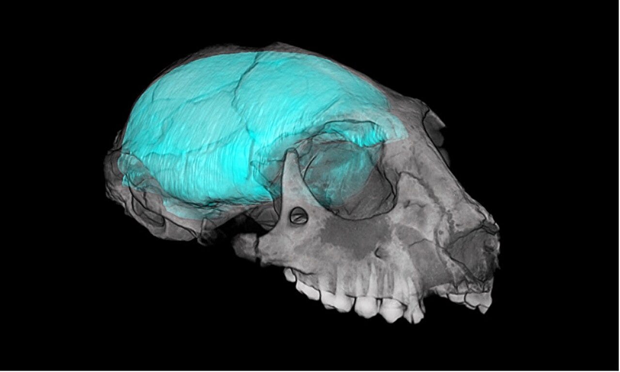
Brains don’t fossilize. That’s the basic problem anyone working on the evolution of primate cognition runs into immediately. Soft tissue rots. What survives is bone, specifically the braincase, and researchers have long used the interior shape of that braincase to infer what the brain inside once looked like. The trouble is that inference from shape alone is subjective. You can look at a fossil skull, notice what seems like a bulging frontal region, and read a story into it that the data don’t actually support.
Kay’s team, working largely from a collection housed at the Duke Lemur Center Museum of Natural History, tried to get around that subjectivity by measuring rather than eyeballing. They micro-CT scanned a wide range of primate skulls, living and extinct, and built digital endocasts, then measured the volumes and surface areas of specific neocortical regions across the primate family tree. The goal was to see which parts of the brain actually grew disproportionately, and where, and when.


